Digital Radiography
Radiography (specifically, x-rays) has long been a valuable tool for diagnosing a range health issues, from broken bones and dental cavities, to pneumonia and concussion syndrome. The digital dental x-ray can be viewed as the logical evolution from analog materials to a computer-based technology that accomplishes the same goal – but does so faster, safer, and for a lower cost.
How Digital Radiography Compares to Film X-Rays
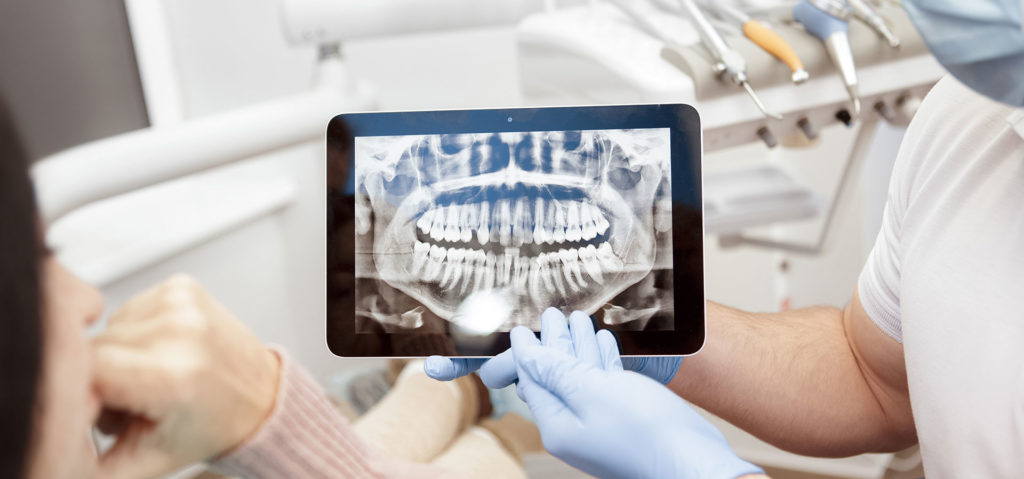
The physical process for digital radiography is actually similar to traditional dental X-rays that use film. Your dentist gently places a sensor into your mouth to capture images of your teeth. But, that’s where the similarities between conventional and digital dental X-rays end. Although it resembles the film used for bitewings and other X-rays, the digital sensor is electronic and connected to a computer. Once the X-ray is taken, the image is projected on a screen for your dentist to view.
From a purely ecological point of view, not only does a digital X-ray greatly reduce material waste, it also doesn’t require the use of potentially harmful chemicals to process images.
Dental X-ray with Less Radiation
The equipment used exposes dental patients to much less radiation. In fact, digital X-rays use up to 90 percent less radiation than film X-rays. While conventional dental X-rays are still relatively safe, digital radiography is an excellent option for those who take X-rays on a regular basis or for those who are concerned about radiation.
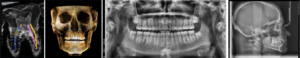
Vatech Green X Digital X Rays
The Green X is an advanced digital X-ray imaging system that incorporates pano, ceph (Optional), CBCT and model scan. It provides high quality images with lower radiation by combining image processing with Vatech’s extensive experience in the dental imaging field. This will improve your diagnostic accuracy, which will lead to increased treatment planning and patient satisfaction.
Large Single Scan FOV
The Green X offers a range of selectable fields of view. The Multi FOV option allows users to select the optimum FOV mode while minimizing exposure to areas that are not in the region of interest. The selection includes the following FOV sizes for diagnostic needs: 18×15, 16×9/16×11, 12×9, 8×8, 8×5, 5×5 and 4×4. These options cover the full arch region, sinus and left/right TMJ, and suits most oral surgery cases and multiple implant surgeries.